|
Hepatobiliary ascariasis is commonly reported from highly endemic regions like India, Bangladesh, Latin America, parts of Middle East and Africa. In humans, the usual habitat of Ascaris lumbricoides is the small intestine. When the worm load is high, going as high as more than 1000 worms, then the worms tend to migrate away from the usual site. Patients with hepatobiliary ascariasis may present with biliary colic due to obstruction caused by the worms in the gall bladder, common bile duct or as a result of obstructive symptoms caused by calcified worms or lithiasis, which is commonly found in patients with hepatobiliary ascariasis. Acute pancreatitis may also be caused by ascariasis. Management usually is conservative if it is still alive or can be extracted by endoscopic retrograde cholangio-pancreatography or surgery.
Keywords: Ascariasis; common bile duct; gallbladder
How to cite this article:
Hajong R. Gall bladder ascariasis. Ann Trop Med Public Health 2013;6:489-90 |
How to cite this URL:
Hajong R. Gall bladder ascariasis. Ann Trop Med Public Health [serial online] 2013 [cited 2020 Sep 23];6:489-90. Available from: https://www.atmph.org/text.asp?2013/6/4/489/127809 |
Ascariasis is mostly seen in tropical and subtropical countries, partly owing to the warm and humid conditions of the soil that is conducive to the development of the ascaris larvae, and partly due to the poor sanitary and hygienic conditions that help in maintaining the infection cycle. [1] The causative organism is Ascaris lumbricoides, which normally lives in the lumen of small intestine. From the intestine, the worm can invade the bile duct or pancreatic duct, but invasion into the gallbladder is quite rare because of the anatomical features of the cystic duct which is narrow and tortuous. [2] Reporting here is a case of biliary ascariasis both gall bladder and common bile duct which was managed by ERCP (endoscopic retrograde cholangio-pancreatography) and laparoscopic cholecystectomy.
A 30-year-old young male presented to the surgery department of NEIGRIHMS Hospital with severe pain in the right upper part of abdomen. The pain was continuous in nature. There was no guarding but tenderness was present over the right hypochondrium region. The patient was icteric. X-ray of abdomen in the erect posture did not reveal any gas under the diaphragm. Ultrasonography was done urgently which showed Ascaris lumbricoides in the gall bladder [Figure 1] and also in the common bile duct [Figure 2]. Total leucocyte count was about 12000/dl and the serum bilirubin was 5.8 mg% with the direct component being higher than the indirect component. Patient did not have fever. Patient was kept nil per orally. Broad spectrum antibiotic was started. Analgesics and antispasmodics were also given. Patient did not get relief of symptoms even after 6 days of conservative management. So Endoscopic retrograde cholangio-pancreatography (ERCP) was done and the common bile duct round worm was extracted [Figure 3] followed by laparoscopic cholecystectomy. The patient recovered uneventfully and was doing well after one year of follow-up postoperatively.
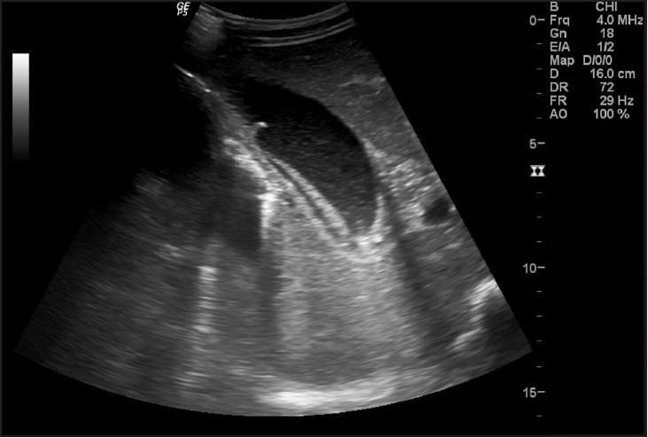 |
Figure 1: Showing ascariasis in gall bladder
Click here to view |
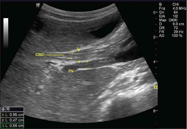 |
Figure 2: Showing CBD ascariasis
Click here to view |
|
Figure 3: Showing CBD worm being extracted by ERCP
Click here to view |
Ascaris lumbricoides has a tendency to migrate through natural body orifices and enter Wirsung’s duct and common bile duct through papilla of Vater. [3] The biliary ascariasis is more common in female. Pregnant women may be more susceptible due to relaxant effect of hormones on the smooth muscle of the bile ducts. The presentation of biliary ascariasis is similar to the cholelithiasis, acute cholecystitis, choledocholithiasis, acute pancreatitis, and ascending cholangitis. The diagnosis of biliary ascariasis usually depends on the demonstration of worms in the biliary tract by different imaging techniques. Sonography has been shown to have a high diagnostic accuracy as a non-invasive procedure in the diagnosis of biliary ascariasis. [4] Various appearances of round worms in the biliary tract and gall bladder have been described. [4],[5] They are as follows:
- Inner tube sign-The round worm may be seen as a thick echogenic stripe with a central anechoic tube (gastrointestinal tract of the worms) in the gall bladder or common bile duct.
- Stripe sign-Thin non-shadowing stripe without an inner tube within gall bladder or common bile duct.
- Spaghettli sign-Overlapping longitudinal interfaces in the main bile duct due to coiling of a single worm or several worms in the common bile duct.
Biliary ascariasis has a good response to conservative treatment like bowel rest, antispasmodic, and anti-helminthic drugs. Worms within the biliary tract are not killed by the anti-helminthic drugs. Successful treatment is possible if the worm returns to the small intestine where they are exposed to adequate concentration of drug. The conservative treatment fails usually in the presence of dead worm, concomitant stones or stricture which prevents the returning of worm in the duodenum. Endoscopic removal of worm treatment has become the treatment of choice for the common bile duct (CBD) ascariasis in which the medical management has failed. Although, biliary ascariasis responds well to medical management but most of the gallbladder ascariasis needs surgical treatment. [6]
| 1. |
Khuroo MS, Zargar SA, Mahajan R. Hepatobiliary and pancreatic ascariasis in India. Lancet 1990;335:1503-6. |
| 2. |
Khuroo MS, Zargar SA, Yatto GN, Dar MY, Javid G, Khan BA, et al. Sonographic findings in gallbladder ascariasis. J Clin Ultrasound 1992;20;587-91. |
| 3. |
Philipe RP, Yune HY. Surgical helminthiasis of the biliary tract. Ann Surg 1960;152;905-10. |
| 4. |
Schulaman A, Loxton AJ, Hegdenrych JJ, Abdul Rahaman KE. Sonographic diagnosis of biliary ascariasis. AJR Am J Roentgenol 1982;139:485-9. |
| 5. |
Cerri GG, Leite GJ, Simoes JB, Correia Da Rocha DJ, Albuquerque FP, Machado MC, et al. Ultrasonographic evaluation of Ascaris in the biliary tract. Radiology 1983;146:753-4. |
| 6. |
Javid G, Wani N, Gulazar GM, Javid O, Khan B, Shah A. Gallbladder ascariasis: Presentation and management. Br J Surg 1999;86;1526-7. |
Source of Support: North Eastern Indira Gandhi Regional Institute of Health and Medical Sciences Hospital, Conflict of Interest: None
DOI: 10.4103/1755-6783.127809
[Figure 1], [Figure 2], [Figure 3] |

