| Abstract |
Dengue viral infection is one of the differential diagnoses in patients having fever with thrombocytopenia. Early diagnosis and timely supportive treatment form the basis for the successful management of these patients. Dengue infection associated with cyanotic congenital cardiac disease makes management more difficult and is associated with a fatal outcome.
We report a case in which the patient presented with fever and thrombocytopenia diagnosed to be a case of dengue infection (NS-1 antigen-positive) and associated cyanotic congenital heart disease (double inlet left ventricle/univentricular heart). The patient was successfully managed with fluids and platelet transfusion.
This case report mainly emphasizes the difficulties faced in hemodynamic monitoring and supportive management during the acute course of the disease.
Keywords: Dengue, double inlet left ventricle, polycythemia, thrombocytopenia
| How to cite this article: Shirodkar CG, Venkategowda PM, Amte R, Rao SM. A rare case of dengue fever in patient with dual inlet left ventricle. Ann Trop Med Public Health 2015;8:37-9 |
| How to cite this URL: Shirodkar CG, Venkategowda PM, Amte R, Rao SM. A rare case of dengue fever in patient with dual inlet left ventricle. Ann Trop Med Public Health [serial online] 2015 [cited 2017 Nov 14];8:37-9. Available from: https://www.atmph.org/text.asp?2015/8/2/37/157286 |
| Introduction |
Dengue fever is caused by a RNA virus that belongs to the family Flaviviridae. Dengue fever is caused by the bite of an infected Aedes mosquito. It is common worldwide: 4 out of 10 people are at risk of this infection, [1] and 100 million new cases have been detected. It causes a wide spectrum of illness, from asymptomatic infection to life-threatening hemorrhagic fever and dengue shock syndrome. [2] Clinical features that are commonly seen are fever, headache, arthralgia, petechial spots, rashes, and hemorrhagic manifestations. A variety of cardiac complications have been reported in dengue-affected patients, such as atrioventricular conduction disorders, supraventricular arrhythmias, and myocarditis.
We report a case of dengue fever with cyanotic congenital heart disease and discuss the implications of cardiac complications in dengue patients. A better understanding of cardiac physiology in congenital heart disease will potentially improve the treatment of dengue illness by avoiding otherwise preventable morbidity and mortality in the affected patients.
| Case report |
A 37-year-old male, a businessman by occupation, presented to our hospital with a history of mild fever and shortness of breath [New York Heart Association (NHYA) grade II] over the past 4 days. He got admitted, initially, to a local hospital where he was diagnosed with thrombocytopenia (with a platelet count 73,000). He was also a known case of cyanotic congenital heart disease (univentricular heart/dual inlet left ventricle with moderate-severe pulmonary stenosis and hypoplastic right ventricle), having been diagnosed in 2007 and on medical management since then. In view of his thrombocytopenia and congenital heart disease, he was referred to our tertiary hospital for further management. At the emergency room (ER) the patient was conscious and coherent. On examination, he was febrile (101°F); with a pulse rate of 78/min, regular, low volume; his blood pressure was 100/60 mmHg with peripheral cyanosis and clubbing (grade 3). Jugular venous pressure (JVP) was elevated, respiratory rate was 25 breaths/min, and SpO 2 was 85% on room air and increased to 91% with face mask (10 L/min O 2 ). [Figure 1] shows the vitals of the patient during admission. Arterial blood gas test (ABG) showed pH 7.42, PCO 2 30.7 mmHg, PaO 2 56.3 mmHg, HCO 3 – 19.6, and SaO 2 89.3%. Cardiovascular system examination showed pansystolic murmur in the left parasternal area, and ejection systolic murmur (ESM) in the pulmonary area with loud P2. Other system examinations were normal. The complete blood count showed hemoglobin 21.6 gm%, packed cell volume (PCV) 64.3 vol%, total leukocyte count 7500/cu mm, and platelet count 9000/cu mm. Coagulation parameters, serum electrolytes, liver function test, and kidney function test were normal. NS-1 antigen for dengue infection was positive, electrocardiogram (ECG) showed sinus rhythm with right axis deviation, chest x-ray showed right paracardiac haziness with straightening of the left heart border [Figure 2]. Two-dimensional echocardiogram (2D echo) showed complex congenital heart disease [single ventricle, double inlet left ventricle, mild aortic valve (AV) regurgitation, moderate-severe pulmonary valve stenosis, normal left ventricular function, and no pericardial effusion]. [Figure 3] shows the univentricular heart/dual inlet left ventricle. Inferior vena cava (IVC) diameter was 2 cm, which is shown in [Figure 4] and [Figure 5]. As the patient had high PCV due to cyanotic congenital heart disease, normal IVC diameter on ultrasound, and single ventricle with moderate-severe pulmonary stenosis, the central venous pressure (CVP) measurement may be insignificant in this case. Fluid resuscitation was done with normal saline according to clinical parameters such as urine output, warm periphery, absence of altered mentation and good pulse volume, and constant monitoring of B line onset on lung ultrasound. The patient received 4 units of platelet-rich plasma for thrombocytopenia only on the first day. The platelet count gradually improved to 20,000 on the second day, and by the third day it was 70,000. Blood culture was sterile. The patient was shifted to a room on the third day and discharged on the fourth day.
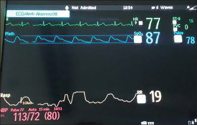 |
Figure 1: Shows vitals during admission of the patient
Click here to view |
| Figure 2: Shows chest x-ray of the patient with right paracardiac haziness
Click here to view |
| Figure 3: Shows dual inlet left ventricle on 2D echo
Click here to view |
| Figure 4: Shows IVC through subcostal approach
Click here to view |
| Figure 5: Shows that there is no collapsibility of IVC during breathing
Click here to view |
| Discussion |
Dengue viral infection is a common cause of fever with thrombocytopenia, occurring worldwide; recent surveillance by the World Health Organization (WHO) shows that its global incidence is increasing. The disease is transmitted through the bite of the mosquito Aedes aegypti, which is found in abundance from latitudes 35° North to 35° South. The clinical features of dengue range from mild fever to multiorgan dysfunction syndrome. The treatment of dengue fever is supportive therapy, which includes platelet concentrate for thrombocytopenia and adequate fluid resuscitation.
Complications of dengue fever are common and usually related to renal and hepatic dysfunction. [3] The association of dengue infection with cardiac complications has been reported. [4],[5] These cardiac complications are relative bradycardia, transient atrioventricular block, and ventricular arrhythmia. [6],[7] The incidence varies greatly from one series to another and may have been underdiagnosed because most of the cases with cardiac complications are clinically mild and self-limited.
Wali et al. reported that 70% of 17 patients with dengue infection suffered diffuse left ventricular hypokinesia, with a mean ejection fraction of 40%, [8] and Kabra et al. reported that 16.7% of 54 children with dengue illness had a decreased left ventricular ejection fraction of <50%. [9] Our patient had congenital cyanotic heart disease, that is, dual inlet left ventricle with pulmonary stenosis. He did not have dengue-related cardiac complications. In view of the mixing of both deoxygenated and oxygenated blood, the ABG showed lower PaO 2 and SaO 2 values. The SpO2 value was 85-91% even with a high-concentration oxygen mask. The SpO 2 did not improve more than 91% for any concentration of oxygen and there was no tachypnea or tachycardia. The patient had polycythemia Hg 21 gm%, and he had a history of phlebotomy 3 years back related to the polycythemia.
Hemodynamic monitoring and fluid resuscitation were the major concerns during the management of our patient. Central venous access and CVP monitoring could not be done, as this patient had thrombocytopenia; the IVC diameter collapsibility index was not useful, as the patient had A single ventricle and valvular disease (pulmonary valve stenosis), and there was high PCV due to underlying chronic cyanosis, but not due to dehydration.
This case report mainly emphasizes the difficulties faced during the management of dengue fever with associated congenital heart disease. Single-ventricle physiology presents a challenge to the intensive care physician because patients with such conditions respond differently to interventions such as supplemental oxygen, mechanical ventilation, fluids, and vasopressors from patients with conventional circulations. Good coordination is required between the cardiologist and the intensive care physician to treat the complications of congenital heart disease and its implication in the treatment of dengue infection to prevent morbidity and mortality.
| Acknowledgments |
We gratefully acknowledge the physician, cardiologist, nurses, and management of the hospital for their valuable support.
| References |
| 1. |
World Health Organization. Dengue Hemorrhagic Fever: Diagnosis, Treatment, Prevention and Control. Geneva: World Health Organization (WHO); 1997. p. 1-84.
|
| 2. |
Koley TK, Jain S, Sharma H, Kumar S, Mishra S, Gupta MD, et al. Dengue encephalitis. J Assoc Physicians India 2003;51:422-3.
|
| 3. |
Monath TP. Dengue: The risk to developed and developing countries. Proc Natl Acad Sci USA 1994;91:2395-400.
|
| 4. |
Satarasinghe RL, Arultnithy K, Amerasena NL, Bulugahapitiya U, Sahayam DV. Asymptomatic myocardial involvement in acute dengue virus infection in a cohort of adult Sri Lankans admitted to a tertiary referral centre. Br J Cardiol 2007;14:171-3.
|
| 5. |
Kularatne SA, Pathirage MM, Kumarasiri PV, Gunasena S, Mahindawanse SI. Cardiac complications of a dengue fever outbreak in Sri Lanka, 2005. Trans R Soc Trop Med Hyg 2007;101:804-8.
|
| 6. |
Chuah SK. Transient ventricular arrhythmia as a cardiac manifestation in dengue haemorrhagic fever–a case report. Singapore Med J 1987;28:569-72.
|
| 7. |
Lateef A, Fisher DA, Tambyah PA. Dengue and relative bradycardia. Emerg Infect Dis 2007;13:650-1.
|
| 8. |
Wali JP, Biswas A, Chandra S, Malhotra A, Aggarwal P, Handa R, et al. Cardiac involvement in dengue haemorrhagic fever. Int J Cardiol 1998;64:31-6.
|
| 9. |
Kabra SK, Juneja R, Madhulika, Jain Y, Singhal T, Dar L, et al. Myocardial dysfunction in children with dengue haemorrhagic fever. Natl Med J India 1998;11:59-61.
|
Source of Support: None, Conflict of Interest: None
| Check |
DOI: 10.4103/1755-6783.157286
| Figures |
[Figure 1], [Figure 2], [Figure 3], [Figure 4], [Figure 5]

